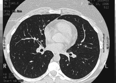
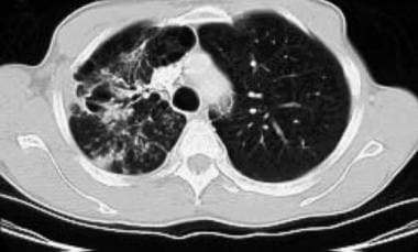
tree in bud radiology assistant
CT provides accurate diagnosis in pulmonary TB in 91 of patients and correctly excludes it in 76 of patients 4. 1 those with a tree-in-bud appearance and 2 those with ill-defined centrilobular nodules of GGA without a tree-in-bud appearance.

Tree In Bud In Centrilobular Nodules The Recognition Grepmed
In humans a CT treeinbud pattern has been described as a characteristic of centrilobular bronchiolar dilation with bronchiolar plugging by mucus pus or fluid.
. Tree-in-bud pattern seen on high-resolution CT HRCT indicates dilatation of bronchioles and their filling by mucus pus or fluid. Along subpleural surface and fissures along interlobular septa and the peribronchovascular bundle. Access consultant level radiologists at any hour of the day all year round.
Aims of this retrospective descriptive multicenter study were to characterize the CT appearance of a treeinbud pattern in a group of cats and compare this pattern with radiographic and clinical. The Department of Radiology Drs. Lung-RADS Lung Imaging Reporting and Data System is a classification proposed to aid with findings in low-dose CT screening exams for lung cancerThe goal of the classification system is to standardize follow-up and management decisions.
CTHRCT are particularly helpful in the detection of small foci of cavitation tree-in-bud pattern and in pleural evaluation namely tuberculous effusion empyema and bronchopleural fistula. Tree-in-bud refers to a pattern seen on thin-section chest CT in which centrilobular bronchial dilatation and filling by mucus pus or fluid resembles a budding tree. 78 indicating the absenceresolution of TIB opacities 26 incomplete thoracic CT scan studies 75 duplicate.
Or have a so-called tree-in-bud appearance Fig 49 Additionally nodules may ei-ther be calcified as occurs in fungal disease or cavitary as is seen for example in patients with. Tree-in-bud almost always indicates the presence of. A 50 years old lady presented with fever productive cough and occasional haemoptysis for two weeks.
Revision requested December 10. The Tree-in-Bud Sign. However the most common process leading to this CT appearance is infection.
Of Medicine Medical College 88 College Street Kolkata 700 073. The tree-in-bud appearance was characterised by well-defined centrilobular nodules of soft-tissue attenuation that were connected to linear and branching opacities. The system is similar to the Fleischner criteria but designed for the subset of patients intended for low-dose.
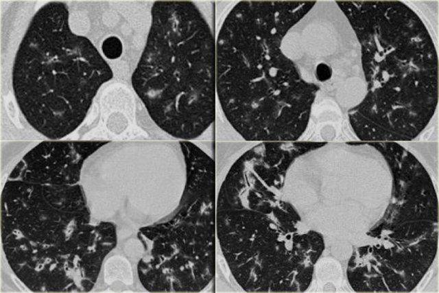
Tree-in-bud refers to a pattern seen on thin-section chest CT in which centrilobular bronchial dilatation and filling by mucus pus or fluid resembles a budding tree Usually somewhat nodular in appearance the tree-in-bud pattern is generally most pronounced in the lung periphery and associated with abnormalities of the larger airways. Basic interpretation Robin Smithuis Otto van Delden and Cornelia Schaefer-Prokop Radiology Department of the Rijnland Hospital Leiderdorp and the Academical Medical Centre Amsterdam the Netherlands Secondary lobule Reticular pattern Nodular pattern Algorithm for nodular pattern Tree-in-bud. The Radiology Assistant HRCT part I.
High-resolution CT usually reveals small 24-mm centrilobular nodules and branching linear opacities of similar caliber originating from a single stalk Figs 2 3 4. Small nodules in a perilymphatic distribution ie. This pattern is most pronounced in the lung periphery and is usually associated with abnormalities of the larger.
Of these 182 cases were excluded for the following reasons. Usually somewhat nodular in appearance the tree-in-bud pattern is generally most pronounced in the lung periphery and associated with abnormalities of the larger airways. Tree in bud opacification refers to a sign on chest CT where small centrilobular nodules and corresponding small branches simulate the appearance of the end of a branch belonging to a tree that is in bud.
The tree-in-bud pattern indicates disease affecting the small airways. Our PETCT reporting services allow Specialty Teleradiology to assist your physicians in. Provide a broader level of service with support from leading sub-specialist radiologists.
Despite atypical findings for COVID-19 pneumonia RT-PCR test was positive for COVID-19. 1 Department of Radiology Graduate Medicine Education University of Wisconsin Medical School and Hospital and Clinics Madison 53792-3252. Revision received and accepted May 22 2000.
Upper and middle zone predominance. 1 From the Department of Radiology University of Vienna Waehringer Guertel 18-20 A-1090 Vienna Austria. Address correspondence to the author e-mail.
Our Radiology Information System was searched for the term tree-in-bud from January 1 2010 to December 31 2010 iden-tifying 599 examinations. Endobronchial spread of infection TB MAC any bacterial bronchopneumonia Airway disease associated with infection cystic fibrosis bronchiectasis less often an airway disease associated primarily with mucus retention allergic bronchopulmonary aspergillosis asthma. Differential diagnosis is broad which includes different etiologies.
Vlahos and Naidich Tisch Hospital New York University Medical Center. Thus the bronchioles resemble a branching or budding tree and are usually somewhat nodular in appearance. The centrilobular nodules were divided into two patterns.
Received November 11 1999. Thin section CT shows peribronchial thickening and centrilobular nodules with tree in bud appearance. The tree-in-bud pattern represents centrilobular branching structures that resemble a budding tree.
She also had generalized weakness and shortness of breath for same duration. The pattern reflects a spectrum of endo- and peribronchiolar disorders including mucoid impaction inflammation andor fibrosis 154. Lymphadenopathy in left hilus right hilus and paratracheal 1-2-3 sign.
The differential diagnosis is lengthy. The tree-in-bud pattern occurs commonly in patients with endobronchial spread of Mycobacterium tuberculosis and is highly suggestive of active tuberculosis 2 3.

The Radiology Assistant Hrct Common Diagnoses
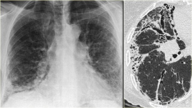
The Radiology Assistant Hrct Common Diagnoses
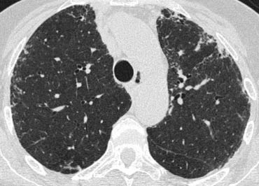
The Radiology Assistant Hrct Basic Interpretation

Learningradiology Lung Abscess Pulmonary Lunges Pulmonary X Ray

Csfoma Peritoneal Cerebrospinal Fluid Pseudocyst Csf Shunt Complication Xray X Ray Student Medical Radiology Imaging Radiology Radiologic Technology
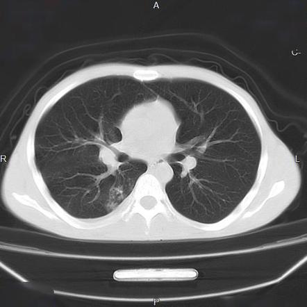
Tree In Bud Sign Lung Radiology Reference Article Radiopaedia Org

The Radiology Assistant Cystic Lung Cancer
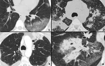
The Radiology Assistant Hrct Basic Interpretation

Case 24 2001 A 46 Year Old Woman With Chronic Sinusitis Pulmonary Nodules And Hemoptysis Nejm
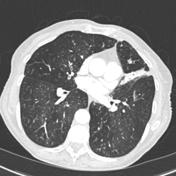
Tree In Bud Sign Lung Radiology Reference Article Radiopaedia Org
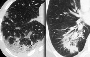
The Radiology Assistant Hrct Basic Interpretation

Air Trapping Radiology Reference Article Radiopaedia Org
The Radiology Assistant Hrct Common Diagnoses

Reverse 3 And 3 Sign Coarctation Abstract Artwork Antonio Mora Artwork Artwork
